Image Segmentation of Pelvic Floor Features & Slings/Meshes
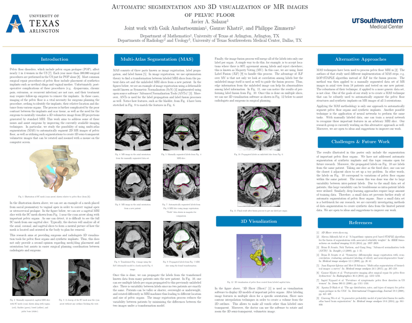
 Pelvic floor disorders, which include pelvic organ prolapse, affect nearly 1 in 4 women in the US. Each year more than 300,000 surgical procedures are performed in the US to treat these disorders. Most common surgical repair procedures of pelvic floor include placement of synthetics implants such as urethral slings and vaginal meshes. Unfortunately, postoperative complications of these procedures (e.g. dyspareunia, chronic pain, extrusion, or recurrent infection) are not rare, and their treatment may require follow-up surgeries to remove the implants. In these cases, imaging of the pelvic floor is a vital necessity for surgeons planning the procedure, seeking to identify the implants, their relative location and distance from various organs. The process is further complicated by the poor contrast between the implants and scar tissue, as well as the need for the surgeons to mentally generate a 3D volumetric image from 2D projections generated by standard MRI. This work aims to address some of these issues and assist surgeons by improving the currently available imaging techniques. In particular, we study the possibility of using multi-atlas segmentation (MAS) to automatically segment 2D MR images of pelvic floor, as well as utilizing such segmentations to create 3D semi-transparent volumetric images that can be rotated and zoomed with a mouse on the computer screen. Press blue link above for poster PDF.

